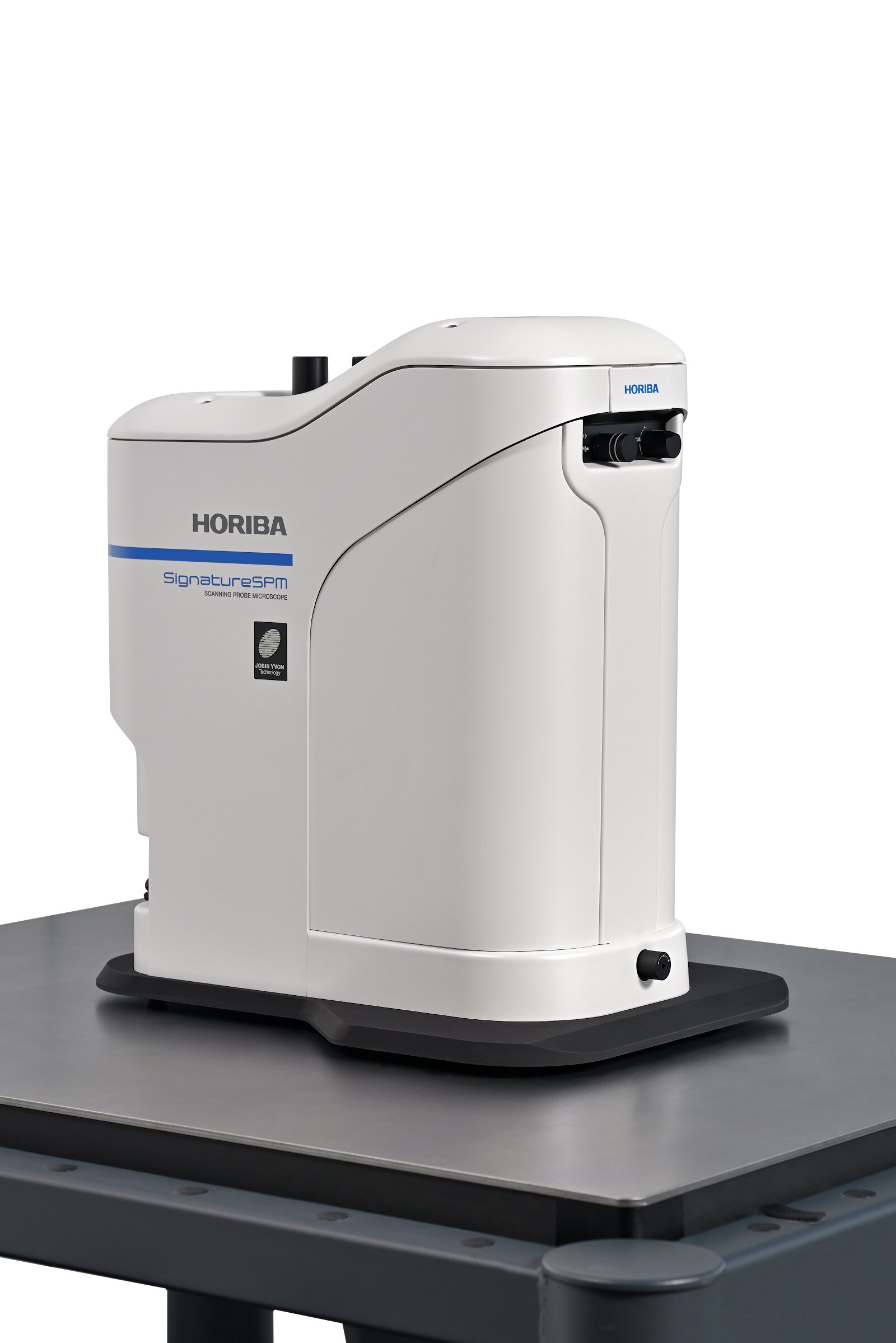
The Molecular Vista SignatureSPM is a scanning probe microscope that delivers simultaneous AFM and nanoscale IR/Raman spectroscopy via Photo-induced Force Microscopy (PiFM), providing chemical imaging with approximately 10 nm spatial resolution — far below the optical diffraction limit — for materials characterization at the nanoscale.
The Molecular Vista SignatureSPM is an advanced scanning probe microscopy platform built around Photo-induced Force Microscopy (PiFM), a proprietary technique that enables simultaneous atomic force microscopy (AFM) topography mapping and infrared or Raman nanospectroscopy at a spatial resolution of approximately 10 nm. By detecting photoinduced force between a sharp AFM tip and the sample surface, PiFM achieves chemical identification and mapping entirely independent of the optical diffraction limit, accessing length scales inaccessible to conventional confocal Raman or FTIR microscopy.
In PiFM mode, an infrared or Raman laser is focused at the tip-sample junction. The electromagnetic field enhancement at the sharp metallic tip drives a photoinduced force that is detected by the AFM cantilever. Because the measurement is force-based rather than photon-based, the spatial resolution is determined by the tip radius (typically 10–20 nm) rather than the wavelength of light. This allows the SignatureSPM to generate full IR or Raman spectra and 2D chemical maps at true nanoscale resolution from a single acquisition, with spectral quality sufficient for chemical identification and library matching.
The SignatureSPM integrates seamlessly with HORIBA Raman excitation sources and spectrographs, providing a complete Nano-Raman/PiFM solution. A key advantage of PiFM over tip-enhanced Raman spectroscopy (TERS) is that it works in ambient conditions without requiring complex tip preparation or vacuum, making it significantly more accessible for routine laboratory use. Samples can be measured in air at room temperature, and the non-contact, non-destructive nature of the measurement preserves delicate soft materials including polymers, biological specimens, and organic thin films.
Typical applications include nanoscale morphology and chemical domain mapping in block copolymers and polymer blends, identification of contaminants and inclusions in semiconductor or electronic materials, characterization of 2D materials and heterostructures at the flake level, protein aggregation and biological membrane studies, and nanoscale defect analysis in organic photovoltaics. NewRoad provides the Molecular Vista SignatureSPM in Israel for materials science, semiconductor, and life science research groups requiring nanoscale chemical imaging beyond the optical diffraction limit.
WHY CHOOSE THIS PRODUCT
Photo-induced Force Microscopy (PiFM) — unique technique combining AFM and nanoscale IR/Raman spectroscopy with ~10 nm spatial resolution
Below the optical diffraction limit — chemical imaging at length scales inaccessible to confocal Raman, FTIR, or TERS microscopy
Simultaneous topography and chemistry — co-registered AFM surface map and hyperspectral chemical image in a single acquisition
Ambient operation — no vacuum, no special tip preparation; works in air at room temperature on soft and hard materials
Broad application range — polymers, semiconductors, 2D materials, biological samples, organic electronics, nanoscale defect analysis
Specifications & Performance
Type
Scanning probe microscope with integrated Photo-induced Force Microscopy (PiFM) for nanoscale IR and Raman spectroscopy
Spatial resolution
~10 nm chemical imaging spatial resolution — far below the optical diffraction limit
Technique
PiFM: force-based detection of photoinduced tip-sample interaction; simultaneous AFM topography and chemical mapping
Operation
Ambient conditions (air, room temperature); non-contact, non-destructive; no vacuum or special tip preparation required
Surface science and nanoscale measurement instruments support life science researchers in studying biomolecular interactions, cell adhesion, and membrane behavior with high precision.
Spectroscopy
Chemistry & Biochemistry
Versatile analytical and surface characterization instruments support fundamental and applied research in molecular chemistry, reaction kinetics, colloid science, and biomolecular analysis.
Surface Science
Cosmetics
Objective surface measurement tools help cosmetic scientists quantify spreadability, adhesion, wettability, and film formation — driving faster, data-driven formulation development.
Electrochemistry
Electrochemistry
Precision electrochemical instruments enable fundamental research and applied development in batteries, sensors, corrosion science, electrodeposition, and electrocatalysis.
Electrochemistry
Energy
Advanced electrochemical and surface characterization tools accelerate research and development of batteries, fuel cells, supercapacitors, and photovoltaic systems.
Material Science
Materials Science
A comprehensive suite of surface, electrochemical, and structural characterization instruments enables materials scientists to understand, optimize, and validate the properties of advanced materials.
Spectroscopy
Pharmaceutical
Pharmaceutical R&D and manufacturing rely on surface analysis, compound characterization, and real-time interface monitoring to develop safe, effective, and consistently produced drug products.
Nanotechnology
Semiconductors
High-sensitivity surface and thin-film characterization instruments are essential for semiconductor wafer processing, yield optimization, and failure analysis at the nanoscale.
RELATED PRODUCTS
Explore Similar Solutions
Browse related instruments and complementary solutions selected for your research and application.